Anatomy Scan Down Syndrome
Anatomy scan down syndrome. Ultrasound can be very helpful in refining the risk calculations. Our midwives wanted us to do it with a specialist because of my history with pre-eclampsia with Emma. If the scan shows that your baby is growing too quickly or too slowly extra scans may be offered to check your babys growth.
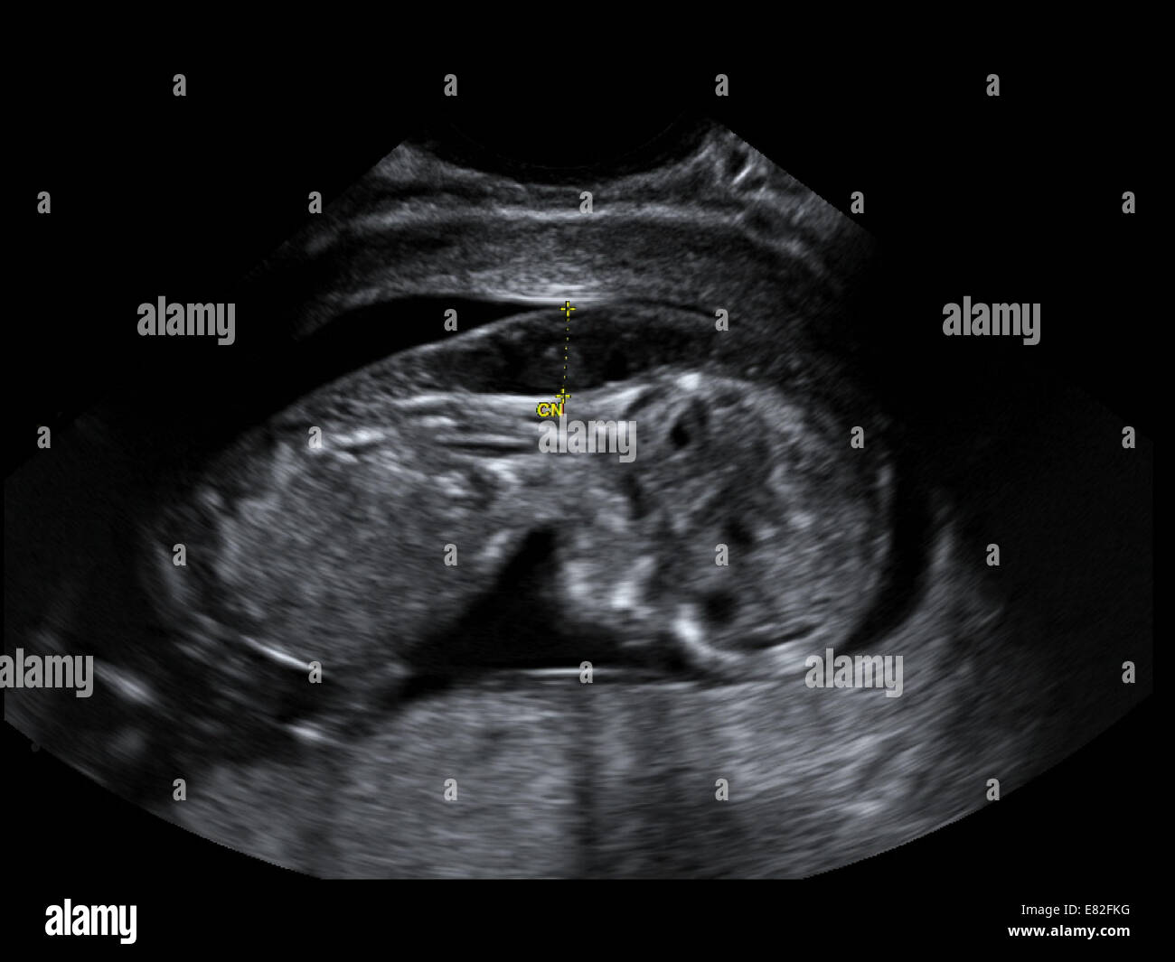
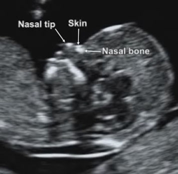
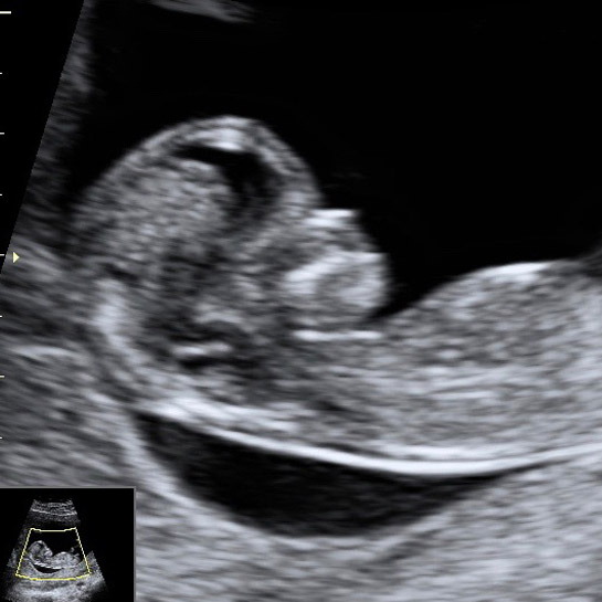
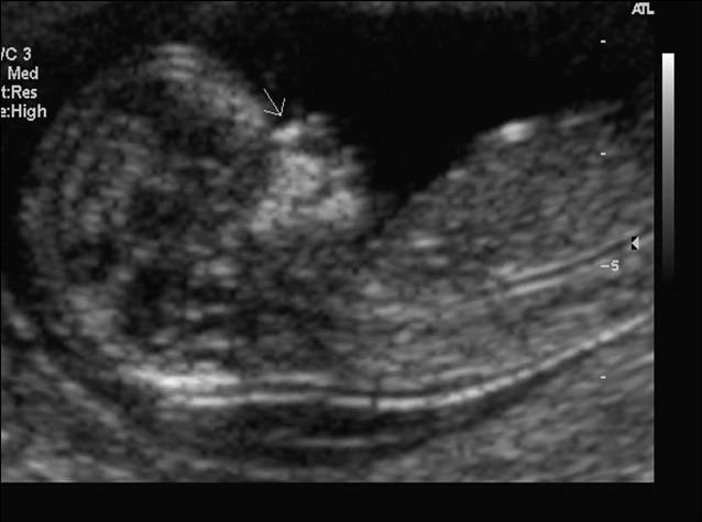
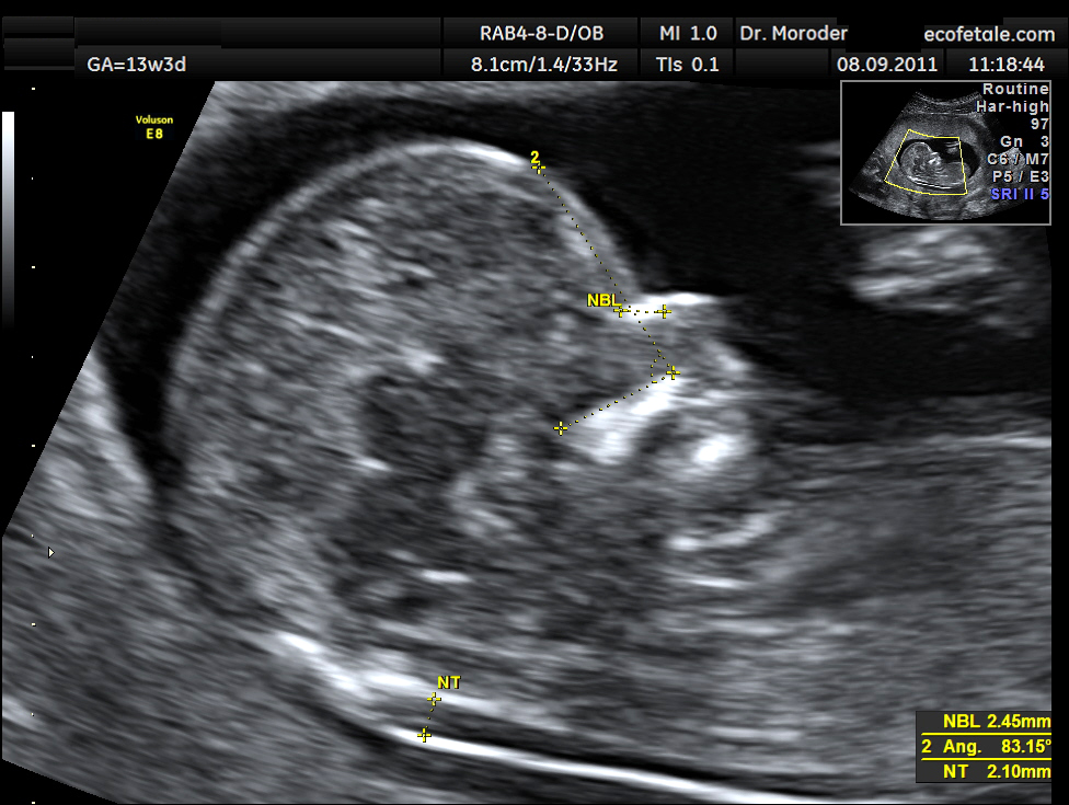
One soft marker that might have shown up on the first-trimester NT screening which is always performed between weeks 10 and 13 is nuchal-fold thickening where the area at the back of a babys neck accumulates fluid causing it to appear thicker than usual. From the scan I cannot see any indicators to indicate Down Syndrome although I am as sure as I can be but can not be 100 That was enough for me. What is Down syndrome.
We then went for an ultrasound and all the babys measurements were normal besides a dilated kidney and echogenic focus bright spot on the heart. Ultrasound markers that can be detected in the second trimester of pregnancy are strongly predictive for Downs syndrome show findings from a systematic review. Neuroanatomy of Downs syndrome.
I cried I felt so relieved. Down syndrome markers at anatomy scan. The results largely confirm findings of previous studies with respect to overall patterns of brain volumes in Downs syndrome and also provide new evidence for abnormal volumes of specific regional tissue components.
Most doctors do an ultrasound early in the second trimester between 16 and 20 weeks. The blood test that is used to screen for Downs syndrome is usually taken at the time of the nuchal translucency scan but the research data suggests that the results are actually more accurate if the blood is taken at 9 weeks rather than 12 weeks. The anatomy scan is offered when you are 1820 weeks pregnant.
This test is however only 991 accurate and an amnio is recommended following the blood test if results are positive. If they are found you will be told that there is a high risk that your baby has Downs and you may be offered more invasive testing - namely an amniocentesis or chorionic villus sampling CVS test. Some health problems do not show up on the fetal anatomy scan.
We will have one more ultrasound in two weeks to confirm. Having a scan is usually a happy event but remember that sometimes scans.
Ultrasound can be very helpful in refining the risk calculations.
Parts of your babys body will be measured to check that they are growing as expected and to look for any problems. Soft markers are sonographic findings that do not in themselves cause any adverse outcomes. Neuroanatomy of Downs syndrome. Cells seem to tolerate this better than having. If they are found you will be told that there is a high risk that your baby has Downs and you may be offered more invasive testing - namely an amniocentesis or chorionic villus sampling CVS test. They found a heart defect and shorter humerus and femur bones. Most doctors do an ultrasound early in the second trimester between 16 and 20 weeks. This test is however only 991 accurate and an amnio is recommended following the blood test if results are positive. Having an extra copy of this chromosome means that each gene may be producing more protein product than normal.
This soft marker has a higher correlation to Down syndrome than any other. Certain second trimester markers for Downs syndrome that are identified in an ultrasound are more significant than others. Ultrasound can be very helpful in refining the risk calculations. I cried I felt so relieved. Down syndrome is a developmental disorder caused by an extra copy of chromosome 21 which is why the disorder is also called trisomy 21. The first one found an echogenic focus in the heart which is what lead us to the NIPT. Down syndrome markers at anatomy scan.



/babyboyultrasound-7bf2ced4b4794754b67dea974b7ec744.jpg)











Post a Comment for "Anatomy Scan Down Syndrome"